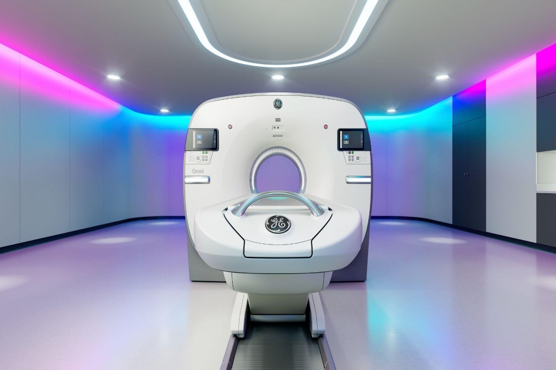
Advancements and Applications in PET-CT Scanners and PET Imaging Technologies

In recent years, positron emission tomography (PET) and computed tomography (CT) have emerged as powerful imaging modalities in the field of medical diagnostics. PET-CT scanners, which combine the functional information from PET with the anatomical details from CT, have revolutionized the way physicians diagnose and manage various diseases. This article explores the recent advancements and applications of PET-CT scanners and PET imaging technologies, highlighting their impact on healthcare and patient outcomes.
Introduction to PET-CT Scanners and PET Imaging Technologies
PET imaging is a nuclear medicine technique that uses radioactive tracers to visualize and measure metabolic processes in the body. It is particularly useful in oncology, cardiology, and neurology, where it can provide valuable insights into the physiological functions of tissues and organs. CT, on the other hand, is a diagnostic imaging technique that uses X-rays to create detailed cross-sectional images of the body’s internal structures.
PET-CT scanners combine the functional information obtained from PET with the anatomical details provided by CT, allowing physicians to pinpoint the location of abnormalities with greater accuracy. This hybrid imaging modality has significantly improved the detection and characterization of various diseases, leading to more precise diagnoses and treatment planning.
Advancements in PET-CT Scanners and PET Imaging Technologies
Recent advancements in PET-CT scanners and PET imaging technologies have focused on improving image quality, reducing radiation dose exposure, and enhancing the efficiency of data acquisition and processing. Manufacturers have introduced new detector technologies, such as time-of-flight (TOF) and silicon photomultiplier (SiPM) detectors, which improve the spatial and temporal resolution of PET images.
Furthermore, advancements in software algorithms have enabled more accurate image reconstruction and quantitative analysis of PET data. This has led to the development of new PET tracers that target specific biological processes, such as tumor metabolism or neuroreceptor binding, opening up new avenues for personalized medicine and targeted therapies.
Applications of PET-CT Scanners and PET Imaging Technologies
The applications of PET-CT scanners and PET imaging technologies span a wide range of medical specialties, with oncology being one of the most prominent areas of use. PET-CT is widely used in cancer staging, treatment response assessment, and surveillance, allowing oncologists to tailor treatment plans based on the metabolic activity of tumors.
In cardiology, PET-CT can assess myocardial perfusion and viability, helping cardiologists diagnose coronary artery disease and plan interventions such as angioplasty or bypass surgery. PET-CT is also valuable in neurology, where it can detect changes in brain metabolism associated with neurodegenerative diseases like Alzheimer’s or Parkinson’s disease.
In addition to clinical applications, PET-CT scanners and PET imaging technologies play a crucial role in research and drug development. PET imaging is used in preclinical studies to evaluate the pharmacokinetics and pharmacodynamics of new drugs, providing valuable insights into their efficacy and safety profiles.
Future Directions and Conclusion
The future of PET-CT scanners and PET imaging technologies looks promising, with ongoing research focusing on further improving image quality, developing new tracers, and expanding the applications of PET-CT in areas such as inflammation imaging and infectious disease diagnostics. As these technologies continue to evolve, they are expected to have an even greater impact on healthcare by enabling earlier and more accurate diagnoses, leading to improved patient outcomes and reduced healthcare costs.
